General & Cosmetic Dentistry
The office of Keith A. Kye, DDS, FAGD serves the neighborhoods of Huntersville, Lake Norman, Davidson and Cornelius.


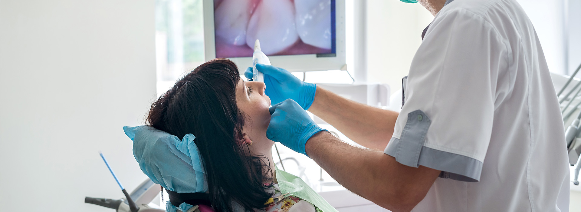
An intraoral camera is a compact, pen-sized imaging device designed to capture clear, full-color views from inside the mouth. Unlike traditional mirrors or handheld inspection tools, the camera provides magnified, high-resolution images of teeth, gums, restorations, and soft tissues. These images reveal surface texture, cracks, early decay, plaque accumulation, and subtle changes in soft tissue that can be difficult to detect with the naked eye alone.
Because the camera feeds in real time to a monitor, clinicians can examine areas from multiple angles and magnifications without repositioning the patient. The visual detail helps identify problems at an earlier stage, which supports more conservative, targeted care. The technology also makes it easier to visually document progression or healing over time, offering a clear point of reference for follow-up visits.
This tool complements other diagnostic aids such as digital X-rays and clinical exams by providing surface-level imaging in color and with immediate visibility. For patients and clinicians alike, the intraoral camera transforms subtle visual information into actionable diagnostic data, making routine exams more thorough and accurate.
One of the most valuable benefits of intraoral imaging is the way it enhances communication. When a patient can see the same image the clinician sees, discussions about oral health become concrete and evidence-based rather than abstract. Visuals reduce uncertainty and allow for clearer explanations of findings, recommended treatments, and preventive strategies.
Showing images during the consultation empowers patients to participate in decisions about their care. It’s easier to explain why a particular area needs attention when a magnified photograph illustrates the issue. This shared view builds trust and helps patients understand the rationale behind treatment priorities and maintenance recommendations.
For the dental team, the intraoral camera streamlines patient education. Rather than relying solely on verbal descriptions or sketches, clinicians can point to specific pixels on the screen—highlighting fractures, margins of restorations, or inflamed tissue—so patients leave the appointment with a precise understanding of their oral condition and next steps.
Intraoral images can be captured and stored as part of a patient’s electronic record, creating a visual timeline of dental health. These photographs are valuable for documenting baseline conditions, tracking changes, and verifying the fit or appearance of restorations over time. Clear, dated images make it easy to compare pre- and post-treatment status during follow-up visits.
Stored images also support continuity of care. When collaboration with specialists or laboratories is necessary, high-quality photographs clarify the clinical situation and streamline communication. Accurate visual records reduce misinterpretation and help outside partners better understand the case before they even examine the patient in person.
From a practice management standpoint, integrating intraoral photos into digital charts improves organization and speeds clinical workflows. With images linked to clinical notes, clinicians can retrieve and reference specific visuals when creating treatment plans, monitoring healing, or responding to clinical questions.
Intraoral cameras play a role in a wide range of clinical tasks. They assist in detecting early caries, identifying cracks or fractures in teeth, evaluating the margins of crowns and fillings, and assessing soft tissue lesions. Because the camera captures nuanced surface detail, it can reveal conditions that might otherwise be missed during a cursory exam.
During restorative and cosmetic consultations, the camera is useful for documenting tooth color, shape, and surface characteristics that inform treatment planning. It helps clinicians and labs make more precise recommendations for veneers, crowns, and other restorations. Post-treatment images verify outcomes and support quality assurance by documenting the finish and fit of work performed.
Clinicians also rely on intraoral imaging to monitor healing after procedures, assess periodontal status, and follow up on suspicious findings. Repeated, comparable images over time are an efficient way to confirm whether tissue is improving, stable, or requires additional intervention.
The intraoral camera exam is quick, comfortable, and noninvasive. During a routine visit, the clinician or hygienist will guide the small camera along the teeth and soft tissues while watching the display. Most patients feel little or no discomfort; the experience is similar to having a routine oral examination, but with the added benefit of visual feedback on a screen.
Patients are encouraged to view the images and ask questions as the clinician points out areas of interest. This collaborative approach helps clarify the nature of any concerns and the reasons behind recommended next steps. Images can be captured at any point during the visit and saved to the patient’s record for future reference.
If further evaluation is needed, the visual findings from the intraoral camera often guide additional diagnostic steps, such as targeted radiographs or a focused clinical assessment. The process is designed to be informative without being intimidating, and clinicians use the images to make the next stage of care as clear and predictable as possible for the patient.
Intraoral cameras have become an indispensable diagnostic and communication tool in modern dental care. By combining precise, real-time imagery with secure digital records, this technology supports earlier detection, clearer patient education, and better coordinated treatment planning. The office of Keith A. Kye, DDS, FAGD employs intraoral imaging as part of a comprehensive approach to oral health, helping patients understand their condition and collaborate on a personalized care plan.
If you’d like to learn more about how intraoral cameras are used in routine exams and advanced care, please contact us for more information.
An intraoral camera is a small, pen-sized imaging device that captures high-resolution, full-color photos and video from inside the mouth. It uses a built-in light source and a high-sensitivity sensor to reveal surface texture, cracks, early decay, plaque, and soft tissue changes that may be difficult to see with the naked eye. Images stream in real time to a monitor so clinicians can examine areas from multiple angles without repositioning the patient.
The camera magnifies details and records them at consistent angles and magnifications to support diagnosis and follow-up. Because it provides immediate visual feedback, the intraoral camera helps clinicians identify problems at an earlier stage and choose more conservative, targeted care. Captured images can also be annotated and saved to the patient record for future comparison and documentation.
Intraoral cameras are useful for spotting a range of surface-level concerns, including early caries, hairline cracks or fractures, worn or failing restorations, and plaque buildup. They also aid in evaluating soft tissue abnormalities such as inflammation, ulcers, or discoloration that warrant closer observation. Because the camera provides magnified color images, subtle differences in texture and margin integrity become easier to identify.
These findings often guide additional diagnostic steps, such as targeted radiographs or focused clinical exams, when deeper assessment is indicated. Repeated images taken over time allow clinicians to monitor progression or healing and determine whether intervention is required. In short, the device extends the clinician's visual capability and improves the chance of catching problems earlier.
Digital X-rays reveal structures beneath the surface, such as tooth roots, bone levels, and interproximal decay, while intraoral cameras document color and surface texture in real time. Together these tools create a more complete diagnostic picture by combining internal and external perspectives of oral health. A clinical exam synthesizes tactile findings with visual evidence from both modalities to support accurate assessment.
Because intraoral images are captured in color and at high magnification, they add a layer of information that X-rays cannot show, such as marginal gaps around restorations or localized soft tissue changes. Clinicians use these complementary data points to refine treatment plans, prioritize care, and provide clearer explanations to patients. The combined documentation also improves recordkeeping and follow-up comparisons.
An intraoral camera exam is generally quick, comfortable, and noninvasive, resembling a routine visual inspection of the mouth. The clinician or hygienist guides the small camera along the teeth and soft tissues while watching the monitor, and most patients feel little or no discomfort during the process. Capturing a series of diagnostic images typically takes only a few minutes within the course of a standard appointment.
Images can be taken selectively at problem areas or comprehensively for baseline documentation, and clinicians will pause to explain notable findings as needed. The brief nature of the exam makes it practical to include intraoral imaging during routine cleanings and evaluations without significantly extending visit length. Patients who prefer to view the images can participate in the discussion in real time.
Intraoral images are captured digitally and stored as part of the patient's electronic dental record, typically with date and descriptive notes for context. These time-stamped images create a visual timeline that clinicians can use to track healing, document the condition of restorations, and compare pre- and post-treatment status. Linking photographs to treatment notes improves organization and speeds retrieval during follow-up visits.
Stored images also support continuity of care when collaboration with specialists or laboratories is needed, since photos convey clinical details that may be hard to describe in words alone. Clinicians rely on consistent, dated imagery to evaluate whether tissue is improving, stable, or requires additional intervention. Proper digital management ensures images remain accessible, organized, and clinically useful over time.
Intraoral cameras transform abstract descriptions into concrete visuals by allowing patients to see the same images their clinician sees on the monitor. This shared view makes it easier to explain findings, demonstrate the extent of an issue, and discuss recommended next steps in plain terms. Visual evidence reduces uncertainty and helps patients make informed decisions about their care.
Clinicians can highlight specific pixels on the image to point out fractures, restoration margins, or areas of inflammation, which enhances patient education during the visit. Seeing clear, magnified photographs also supports realistic expectations for treatment outcomes and follow-up needs. Overall, intraoral imaging streamlines conversations and strengthens the clarity of clinical explanations.
For restorative and cosmetic cases, intraoral cameras document tooth color, shape, and surface characteristics that inform shade selection and design decisions for crowns, veneers, and composite restorations. High-resolution photos help clinicians evaluate margins, contacts, and surface texture before treatment and provide precise references for laboratory communication. Captured imagery also aids in planning esthetic changes by clearly showing baseline conditions.
Post-treatment images are used to verify the fit, finish, and appearance of restorations and to confirm that clinical objectives were met. Consistent photographic documentation supports quality assurance and can guide minor adjustments when needed. By providing objective visual references, intraoral imaging improves coordination between the clinician and dental technicians.
Yes, intraoral images are a valuable tool for collaboration with specialists and dental laboratories because they convey clinical details that are hard to capture in text alone. Photographs clarify the condition of teeth and soft tissues, show restoration margins, and illustrate occlusion and cosmetic concerns that laboratories need to reproduce. Sharing high-quality images speeds communication and reduces the chance of misinterpretation during case planning.
When referring a patient to a specialist, attaching dated intraoral photos to the referral provides a clear visual summary of findings and prior treatments. Laboratories also benefit from detailed images for shade matching and contouring, which can improve the fit and esthetics of prosthetics. Overall, photographic documentation streamlines multidisciplinary care and helps ensure consistent outcomes.
Clinicians follow technique protocols that include proper isolation, consistent angulation, and appropriate lighting to capture reproducible, high-quality images. Using retractors, mirrors, and dry fields when needed helps minimize glare and soft tissue interference, while consistent camera settings and positioning produce comparable shots over time. Training on image capture and documentation standards also improves the clinical value of each photograph.
Images are typically reviewed immediately and, if necessary, retaken from a slightly different angle or magnification to ensure diagnostic clarity. Clinicians annotate or categorize photos in the patient record so images can be referenced efficiently during treatment planning or follow-up. These practices help transform raw photos into reliable clinical tools.
The office of Keith A. Kye, DDS, FAGD uses intraoral cameras to enhance diagnostic accuracy, improve patient education, and maintain thorough visual records that support long-term oral health. Incorporating this imaging technology into routine exams helps clinicians detect subtle surface changes earlier and make more informed decisions about conservative care. Providing clear visuals also helps patients understand conditions and recommended treatment steps in a concrete way.
Using intraoral photography as part of a comprehensive approach to diagnosis and treatment planning promotes better documentation and coordination of care. Patients benefit from consistent, dated images that track healing and restoration performance over time. If you have questions about intraoral imaging or want to see how it is used during an exam, the clinical team can explain the process during your next visit.

The office of Keith A. Kye, DDS, FAGD serves the neighborhoods of Huntersville, Lake Norman, Davidson and Cornelius.